Mycoplasma is a prokaryotic microorganism of the class Mollicutes that lack a true cell wall, and many of which are considered pathogenic. Mycoplasma contamination is often detected in cell cultures, and consequently, virus cultures, vaccines and other biological materials produced in cells also become contaminated.
Mycoplasma contamination in cell lines used for research poses a serious problem. In most cases, visual detection of such contaminations or detection with the aid of a microscope is impossible. Although mycoplasma does not cause visible damage to cells, it undeniably affects cell metabolism, cell growth in culture, protein synthesis, cytokine secretion, and even causes damage to DNA and RNA. Hence, results obtained from experiments are liable to be biased when mycoplasma is present. Various studies show that the percentage of contaminated cultures in cell banks is 10%-80%. Mycoplasma contamination can originate from bovine serum, laboratory employees, other contaminated cultures, or the animals from which the cells have been harvested.
The most prevalent species of mycoplasma detected in contaminated cell cultures include M. fermentans, M. hyorhinis, M. arginini, M. orale, M. salivarium, M. hominis, M. pulmonis and M. pirum.
Mycoplasma Contamination Testing Methods
Several methods for the detection of mycoplasma have been published:
- Cultures on agar, liquid media, or semi-solid media.
- DAPI Staining – staining DNA with fluorescent dyes (4’, 6-diamine-2-phenylindole dihydrochloride).
- DNA hybridization.
- Antibodies for specific mycoplasma species.
- Electronic microscope.
- PCR: specific primers.
The testing required by the regulatory authorities is seeding in culture (agar and liquid media). This test is complicated, time consuming (about 5 weeks), and some mycoplasma species are difficult to detect with this method. In recent years, the disadvantages of these methods have been acknowledged (such as sensitivity, specificity and long and complex procedures), and use of PCR for the detection of contaminations in cell cultures has become increasingly widespread.
Table 1. PCR-Based Metod versus Culture Method
| PCR-BASED METHOD | MICROBIOLOGICAL CULTURE | |
|---|---|---|
| 1 | Rapid; Results within 5 hours | Long and time consuming; Results up to 3 weeks |
| 2 | Simple, one-step reaction; Less than 10 minutes to prepare the sample. The mixture contains everything (Including Taq Poly) | Cumbersome |
| 3 | Can be used routinely in every lab | Requires specialized personnel |
| 4 | Sensitive; *(5-100 CFU/ml) | Sensitive |
| 5 | M. Hyorhinis can be detected | Hard to detect M. Hyorhinis |
*Adequate to diagnose cell cultures infected with mycoplasmas. Infections usually result in mycoplasma titers of 10 5 – 10 8 CFU/ml (Mc Garrily 1982)
Using PCR for the Detection of Mycoplasma
PCR has been shown to be a highly sensitive, specific and rapid method for the detection of mycoplasma contamination in cell cultures. Specific primers have been designed from DNA that is coded to the ribosomal RNA (16SrRNA). The gene sequences for RNA are considered conserved sequences and are similar in the various mycoplasma species, which have not undergone significant mutation. Consequently, primers can be designed for these areas, which are specific to the mycoplasma and will not detect bacterial or animal DNA sequences.
The literature describes several PCR methods for the detection of mycoplasma, such as using a number of primers to obtain detection of specific mycoplasma species, and nested PCR (two consecutive PCR cycles using different primers) for amplifying sensitivity and specificity. PCR testing techniques are based on amplification of a DNA fragment using primers prepared in advance, and fragment identification is usually carried out with electrophoresis.
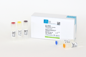
Biological Industries has developed the EZ-PCR Mycoplasma Test Kit, In conjunction with Prof. Shlomo Rottem of the Mycoplasma Laboratory at the Hebrew University-Hadassah Medical School, Jerusalem,
The EZ-PCR Mycoplasma Test Kit is PCR-based and simplifies testing and detection of mycoplasma contamination in cell cultures. The kit includes a unique reaction mix that contains all the ingredients required for PCR: nucleotides, primers, Taq Polymerase and magnesium. No prior preparations are required for PCR, other than the sample to be tested (centrifugation and suspension in the buffer supplied with the kit). After performing agarose gel electrophoresis, positive samples will yield a 270bp fragment. The test takes approximately five hours to complete.
The primers have been designed to detect the mycoplasma species responsible for most contaminations in cell cultures (including Acholeplasma). The primers were tested and found to be specific to mycoplasmatic DNA, and do not react with animal or bacterial DNA.
In sensitivity tests for the detection of defined mycoplasmas, the EZ-PCR Mycoplasma Test Kit was found to be the most sensitive in comparison to other test kits currently available on the market (Table 2). The ability to routinely conduct rapid and simple tests to detect mycoplasma contamination in cell cultures facilitates the eradication or treatment of contaminated cells.
Table 2.Minimal concentration of mycoplasma detected with EZ-PCR Mycoplasma Test Kit
| Mycoplasma specie | Minimal concentration detected |
|---|---|
| M. fermentans | 12.0 CFU/ml |
| M. capricolum | 5.5 CFU/ml |
| M. penetrans | 10.0 CFU/ml |
| M. hyorhinis | 10.5 CFU/ml |
Using Antibiotics to Disinfect Cell Cultures
The increasingly widespread use of more sophisticated and sensitive methods for the detection of mycoplasma contamination in cell cultures has resulted in contamination being detected in numerous cultures. This raises the issue of how to eliminate mycoplasma contamination. Naturally, the ideal solution is to discard the contaminated cells. However, if the cells that are stored in liquid nitrogen are also contaminated, a solution is required for eliminating the mycoplasma and preparing a new cell bank, particularly if the cells are unique and the result of extensive work.
- A number of effective methods for the elimination of mycoplasma contamination in cell cultures have been published, such as:
- Treatment with specific hyperimmune serum (antibodies).
- Passage of contaminated cells in thymus-deficient mice.
- Exposure to analogs of nucleic acids that prevent reproduction of mycoplasma.
- Treatment with antibiotics.
- Exposure of contaminated cells to mouse macrophages.
- A technique that combines growing cells on soft agar and treatment with antibiotics.
The preferred method in terms of simplicity is treatment with antibiotics, which do not damage or alter cells. Antibiotics such as penicillin, which attacks bacterial cell walls, are ineffective in this instance, since mycoplasma lacks a true cell wall. Several antibiotics eliminate mycoplasma effectively, such as: Tylosin, Neomycin, Tetracycline and Gentamicin. However, the efficacy of these antibiotics is restricted to specific mycoplasma species and frequently only reduce the concentration of mycoplasmas rather than disinfect the cell culture. Hence, as soon as treatment is concluded, contamination will recur.
Two methods are recommended for treating contaminated cells with antibiotics. The first is based on alternating treatment with two types of antibiotics, and the second on treatment with one type of antibiotic.
BIOMYC-1 is a solution based on the antibiotic Tiamutin. BIOMYC-2 is a solution based on the antibiotic Minocycline, which is a member of the Tetracycline group.
These two antibiotics have been shown to be effective in eliminating the mycoplasma species frequently present in contaminated cell cultures. Furthermore, the mycoplasmas do not develop resistance to these antibiotics – a common occurrence with other antibiotic treatment methods. The treatment is based on alternating use of the two antibiotics.
BIOMYC-3 is a solution based on the antibiotic Ciprofloxacin. This antibiotic belongs to the Fluoroquinolone group that inhibits synthesis of the enzyme DNA gyrase, which is responsible for supercoiling in bacterial DNA.
Treatment with Ciprofloxacin does not damage or alter the cells. This antibiotic should not be used repeatedly in cell cultures; otherwise, the mycoplasma is liable to develop resistance to it.
Summary
Heightened awareness regarding mycoplasma contamination, and increased use of sensitive and effective methods for the detection and treatment of mycoplasma contaminations, will lead to a reduction in the percentage of contaminated cultures. In addition to isolating contaminated cultures, and discarding or treating them, meticulous work procedures should be followed, and only mycoplasma-free raw materials should be used.