Description
Details
Product Overview
NutriStem® hPSC XF Medium is a widely published, defined, xeno-free, serum-free cell culture medium designed to support the growth and expansion of human induced pluripotent stem (hiPS) and human embryonic stem (hES) cells. Protocols have been established around the world for applications ranging from derivation to differentiation. NutriStem® hPSC XF Medium offers the ability to culture cells in a completely xeno-free medium without the need for high levels of basic FGF and other stimulatory growth factors and cytokines. NutriStem® hPSC XF Medium exhibits a consistent media performance and predictable cellular behavior derived from a defined xeno-free formulation as well as increased reproducibility shown in long-term growth of over 50 passages.
Low-protein formulation that contains stable L-alanyl-L-glutamine and HSA.
NutriStem® hPSC XF Features
- Defined, serum-free, and xeno-free
- Flexible and compatible with multiple matrices
- Amenable to weekend-free culture
- FDA Drug Master File (DMF) available, produced under cGMP
- Enables efficient expansion and growth of hES and hiPS cells in feeder-free culture systems
- Extensively tested and widely used on multiple hES and iPS cell lines, including H1, H9.2, I6, I3.2, and CL1
Sample Data
Cell morphology
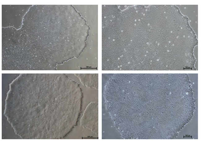
Figure: Normal Colony Morphology. H1 hES cells (top panel) and ACS-1014 hiPS cells (bottom panel) cultured in NutriStem® hPSC XF Medium on Matrigel-coated plates display colony morphologies typical of normal feeder-free hES and hiPS cell cultures, including a uniform colony of tightly compacted cells and distinct colony edges.
Immunostaining
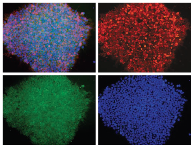
Figure: H1 cell morphology and immunofluorescence analysis of hESC markers red SSEA-4, green OCT4 and blue DAPI. H1 cells stained positive for the expression of pluripotency markers.
Embryoid body formation
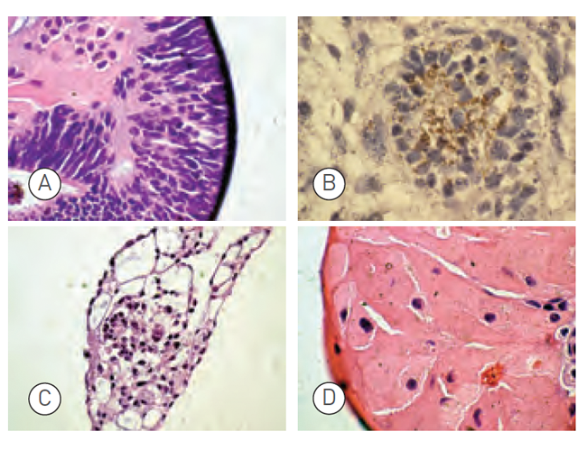
Figure: Embryoid bodies (EBs) were generated from H9.2 hES cells cultured for 16 passages in NutriStem® hPSC XF Medium on Matrigel matrix as an evaluation of pluripotency. The pluripotent H9.2 cells were suspended in serum-supplemented medium, where they spontaneously formed EBs containing cells of embryonic germ layers. The following cell types were identified by examination of the histological sections of 14-day-old EBs stained with H&E: (A) neural rosette (ectoderm), (B) neural rosette stained with Tubulin, (C) primitive blood vessels (mesoderm), and (D) megakaryocytes (mesoderm).
Taratoma formation
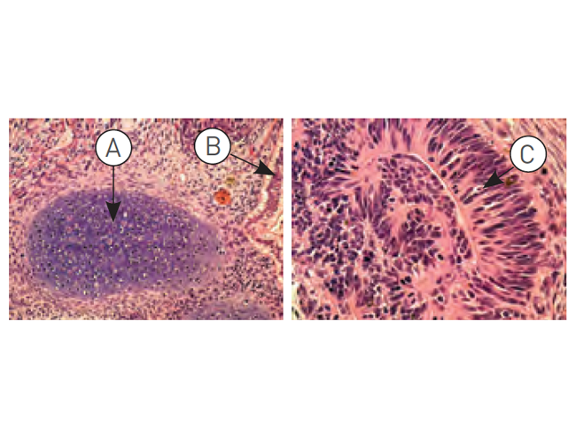
Figure: H9.2 hES cells were cultured for 11 passages in NutriStem® hPSC XF Medium using a human foreskin fibroblast (HFF) feeder layer. The hES cells were subsequently injected into the hind leg muscle of SCID-beige mice for in vitro evaluation of pluripotency. The following tissues from all three germ layers were identified in H&E-stained histological sections of the teratoma 12 weeks post-injection: (A) cartilage (mesoderm), (B) epithelium (endoderm), and (C) neural rosette (ectoderm).
Specifications
Specifications
| Form | Liquid |
|---|---|
| Brand | NutriStem® |
| Storage Conditions | Store at -20ºC |
| Shipping Conditions | Dry Ice |
| Quality Control | NutriStem® hPSC is routinely tested for optimal maintenance and expansion of undifferentiated hESCs. Additional standard evaluations are pH, osmolality, endotoxins and sterility tests. |
| Instructions for Use |
Note: A common feeder-free basement membrane matrix is Matrigel, which is not xeno-free. Effective xeno-free alternatives to Matrigel is recombinant laminin, such as LaminStem(R) 521 (BI Cat. No. 05-753-1F) which has been validated to successfully culture human ES and iPS cells using NutriStem® hPSC XF medium. |
| Legal | A Drug Master File (DMF) for NutriStem® hPSC XF is available. |
References
references
Growing Methods of hESC and iPSC (Derivation, Expansion, Scaling up, and Suspensions)
- O.Thompson,et al. Low rates of mutation in clinical grade human pluripotent stem cells under different culture conditions. Nat Commun 11, 1528 (2020). https://doi.org/10.1038/s41467-020-15271-3
- J. Lee, et al. Induced pluripotency and spontaneous reversal of cellular aging in supercentenarian donor cells. Biochemical and Biophysical Research Communications 27 February 2020, https://doi.org/10.1016/j.bbrc.2020.02.092
- A. Kuwahara, et al. Preconditioning the Initial State of Feeder-free Human Pluripotent Stem Cells Promotes Self-formation of Three-dimensional Retinal Tissue Scientific Reports volume 9, Article number: 18936 (2019)
- A. Keller, et al. Uncovering low-level mosaicism in human embryonic stem cells using high throughput single cell shallow sequencing Scientific Reports, (2019) 9:14844 | https://doi.org/10.1038/s41598-019-51314-6
- I. Uçkay, et al. Regenerative Secretoma of Adipose-Derived Stem Cells from Ischemic Patients Journal of Stem Cell Research & Therapy July 02, 2019 Volume 9 Issue 5
- I. Klimanskaya, Embryonic Stem Cells: Derivation, Properties, and Challenges, Principles of Regenerative Medicine (Third Edition) 2019, chapter 7, pages 113-123
- K. Yoda et al. Optimization of the treatment conditions with glycogen synthase kinase-3 inhibitor towards enhancing the proliferation of human induced pluripotent stem cells while maintaining an undifferentiated state under feeder-free conditions. Journal of Bioscience and Bioengineering, October 2018
- X. Gao et al. A Rapid and Highly Efficient Method for the Isolation, Purification, and Passaging of Human-Induced Pluripotent Stem Cells. Cellular Reprogramming, Vol. 20, No. 5, 2018
- T. Teramura et al. Laser-assisted cell removing (LACR) technology contributes to the purification process of the undifferentiated cell fraction during pluripotent stem cell culture. Biochemical and Biophysical Research Communications, volume 503, Issue 4, 18 September 2018, pages 3114-3120
- Y.Y. Lipsitz et. al. Chemically controlled aggregation of pluripotent stem cells. Biotechnology and Bioengineering, 2018; 1-6
- H. Albalushi et al. Laminin 521 Stabilizes the Pluripotency Expression Pattern of Human Embryonic Stem Cells Initially Derived on Feeder Cells Stem Cells International, Volume 2018
- O.M. Russell et al. Preferential amplification of a human mitochondrial DNA deletion in vitro and in vivo. Scientific Reports, volume 8, Article number: 1799 (2018)
- Maroof M Adil, David V Schaffer. Expansion of human pluripotent stem cells. Current Opinion in Chemical Engineering 2017, 15:24–35
- Tateno, H. et al. Development of a practical sandwich assay to detect human pluripotent stem cells using cell culture media Regenerative Therapy, Volume 6, June 2017, Pages 1–8
- Baker, D. et al. Detecting Genetic Mosaicism in Cultures of Human Pluripotent Stem Cells Stem Cell Reports, 2016
- Vega-Crespo, A., et al. Investigating the functionality of an OCT4-short response element in human induced pluripotent stem cells. Molecular Therapy — Methods & Clinical Development 3, Article number: 16050 (2016)
- Y.Y. Lipsitz, P.W. Zandstra, Human pluripotent stem cell process parameter optimization in a small scale suspensionbioreactor. BMC Proceedings, 9(Suppl 9), O10, 2015
- S. Gregory et al. Autophagic response to cell culture stress in pluripotent stem cells. Biochemical and Biophysical Research Communications, doi:10.1016/j.bbrc.2015.09.080, 2015
- N. Desai, P Rambhia and A. Gishto, Human embryonic stem cell cultivation: historical perspective and evolutionof xeno-free culture systems. Reproductive Biology and Endocrinology 13.1 (2015): 9.
- T. Yokobori et al., Intestinal epithelial culture under an air-liquid interface: a tool for studying human and mouse esophagi. Diseases of the Esophagus, doi: 10.1111/dote.12346. 2015
- L. Healy, L Ruban, Derivation of Induced Pluripotent Stem Cells, Atlas of Human Pluripotent Stem Cells in Culture, pp 149-165. Springer US 2015
- W. Siqin et al. Spider silk for xeno-free long-term self-renewal and differentiation of human pluripotent stem cells. Biomaterials 35.30 (2014): 8496-8502.
- G. Finesilver, M. Kahana, E. Mitrani. Kidney-Specific Micro-Scaffolds and Kidney Derived Serum FreeConditioned Media support in vitro Expansion, Differentiation, and Organization of Human Embryonic Stem Cells. Tissue Engineering Part C: Methods. -Not available-, ahead of print. doi:10.1089/ten.TEC.2013.0574.
- M. Amit, J. Itskovitz-Eldor. Atlas of Human Pluripotent Stem Cells: Derivation and Culturing. Stem Cell Biology and Regenerative Medicine, 2012
- R. Bergström, Xeno-free culture of human pluripotent stem cells, Methods Mol Biol. 2011;767:125-36
- J.Collins et al,. Highly Efficient Reprogramming to Pluripotency and Directed Differentiation of Human Cells withSynthetic Modified mRNA. Cell Stem Cell 7 (5): 618-630 (2010).
- K. Jacobs et al. Higher-Density Culture in Human Embryonic Stem Cells Results in DNA Damage and GenomeInstability. Stem Cell Reports: 6(3), pp 330–341, 2016
Differentiation of Pluripotent Stem Cells
- L. Vitillo et. al. The isochromosome 20q abnormality of pluripotent cells interrupts germ layer differentiation. Stem Cell Reports, vol. 18, pp. 782–797, 2023
- K. Nishimura et. al. A human development-based protocol for the differentiation of human ESCs into midbrain dopaminergic neurons. 2023
- H.G. Tay et al. Photoreceptor laminin drives differentiation of human pluripotent stem cells to photoreceptor progenitors that partially restore retina function. Molecular Therapy, 2023
- F. Cordella et al. Human iPSC-Derived Cortical Neurons Display Homeostatic Plasticity. Life, 2022
- D. Zlotnik et al. P450 oxidoreductase regulates barrier maturation by mediating retinoic acid metabolism in a model of the human BBB. Stem Cell Reports, 2022
- T. Parmentier et al. Human cerebral spheroids undergo 4-aminopyridine-induced, activity associated changes in cellular composition and microrna expression. Scientific Reports, 2022
- L. Wertheim, et al. Regenerating the Injured Spinal Cord at the Chronic Phase by Engineered iPSCs-Derived 3D Neuronal Networks. Adv. Sci. 2022, DOI: 10.1002/advs.202105694
- L. Nazlamova, et al. Generation of a cone photoreceptor specific GNGT2 reporter line in human pluripotent stem cells. Stem Cells, 2022. https://doi.org/10.1093/stmcls/sxab015
- M. Ozgencil, et al. Assessing BRCA1 activity in DNA damage repair using human induced pluripotent stem cells as an approach to assist classification of BRCA1 variants of uncertain significance. PLOS, 2021. https://doi.org/10.1371/journal.pone.0260852
- S. Rajasingh, et al. Comparative analysis of human induced pluripotent stem cell-derived mesenchymal stem cells and umbilical cord mesenchymal stem cells. jcmm, 2021, https://doi.org/10.1111/jcmm.16851
- C. Markouli, et. al. Sustained intrinsic WNT and BMP4 activation impairs hESC differentiation to definitive endoderm and drives the cells towards extra-embryonic mesoderm. Sci Rep 11, 8242 (2021). https://doi.org/10.1038/s41598-021-87547-7
- Á. P. Reyes, Developmental Insights and Biomedical Potential of Human Embryonic Stem Cells : Modelling Trophoblast Differentiation and Establishing Novel Cell Therapies for Age-related Macular Degeneration. Karolinska Institutet (Sweden), ProQuest Dissertations Publishing, 2020. 28420975.
- R. Schick, et al. Electrophysiologic Characterization of Developing Human Embryonic Stem Cell-Derived Photoreceptor Precursors. Investigative Ophthalmology & Visual Science September 2020, Vol.61, 44. doi:https://doi.org/10.1167/iovs.61.11.44
- P. Sunderland, et al. ATM-deficient neural precursors develop senescence phenotype with disturbances in autophagy. Mechanisms of Ageing and Development.
1 July 2020, https://doi.org/10.1016/j.mad.2020.111296 - S. P. Sanchez, SCALABLE, SAFE AND GMPCOMPATIBLE PRODUCTION OF EMBRYONIC STEM CELL DERIVED RETINAL PIGMENT EPITHELIAL CELLS. Karolinska Institutet, Stockholm, Sweden, March 27th, 2020
- L. Tolosa et al., Transplantation of hESC-derived hepatocytes protects mice from liver injury Stem Cell Research and Therapy, BioMed Central, 2015, 6 (1), pp.246. ff10.1186/s13287-015-0227-6ff. ffinserm-01254139f
- A. Shoval, et al. Anti‐VEGF‐Aptamer Modified C‐Dots—A Hybrid Nanocomposite for Topical Treatment of Ocular Vascular Disorders Wiley Online Library, 12 August 2019 https://doi.org/10.1002/smll.201902776
- Y. Chemla et al., Gold nanoparticles for multimodal high-resolution imaging of transplanted cells for retinal replacement therapy NANOMEDICINE, VOL. 14, NO. 14, 24 Jul 2019, https://doi.org/10.2217/nnm-2018-0299
- P.Ni et al. iPSC-derived homogeneous populations of developing schizophrenia cortical interneurons have compromised mitochondrial function Molecular Psychiatry, 31 July 2019
- L. P. Liu et al. Therapeutic Potential of Patient iPSC-Derived iMelanocytes in Autologous Transplantation Volume 27, Issue 2, Cell Reports, 9 April 2019, Pages 455-466.e5
- C. M. Sellgren et al. Increased synapse elimination by microglia in schizophrenia patient-derived models of synaptic pruning. Nature Neurosciencevolume 22, pages374–385 (2019)
- S. Su et al. A Renewable Source of Human Beige Adipocytes for Development of Therapies to Treat Metabolic Syndrome. Cell Reports, Volume 25, Issue 11, Pages 2935-3230, 2018
- SL Ji and SB Tang, Differentiation of retinal ganglion cells from induced pluripotent stem cells: a review. Int J Ophthalmol. 2019; 12(1): 152–160.
- A. MarKus et al. An optimized protocol for generating labeled and transplantable photoreceptor precursors from human embryonic stem cells. Experimental Eye Research, volume 180, March 2019, pages 29-38
- Z. Shao et al. Dysregulated protocadherin-pathway activity as an intrinsic defect in induced pluripotent stem cell–derived cortical interneurons from subjects with schizophrenia. Nature Neuroscience volume 22, pages 229–242 (2019)
- M. Tewary, Engineered In vitro models of post-implantation human development to elucidate mechanisms of self-organized fate specification during embryogenesis. A thesis submitted, Institute of Biomaterials and Biomedical Engineering, University of Toronto, 2018
- D.L. McPhie et al. Oligodendrocyte differentiation of induced pluripotent stem cells derived from subjects with schizophrenias implicate abnormalities in development Translational Psychiatryvolume 8, Article number: 230 (2018)
- J. Ameri et al. Efficient Generation of Glucose-Responsive Beta Cells from Isolated GP2+ Human Pancreatic ProgenitorsCell Reports, Volume 19
- R. De santis et al. Direct conversion of human pluripotent stem cells into cranial motor neurons using a piggyBac vector. Stem Cell Research 29 (2018) 189–196
- K.M. Gray et al. Self-oligomerization regulates stability of Survival Motor Neuron (SMN) protein isoforms by sequestering an SCFSlmb degron. Molecular Biology of the Cell, 2017 mbc.E17-11-0627
- E. Welby et al. Isolation and Comparative Transcriptome Analysis of Human Fetal and iPSC-Derived Cone Photoreceptor Cells. Stem Cell Reports (2017), https://doi.org/10.1016/j.stemcr.2017.10.018
- R. De-Santis et. al. FUS Mutant Human Motoneurons Display Altered Transcriptome and microRNA Pathways with Implications for ALS Pathogenesis. Stem Cell Reports (2017), https://doi.org/10.1016/j.stemcr.2017.09.004
- R.A. Hazim et al. Differentiation of RPE cells from integration-free iPS cells and their cell biological characterization. Stem Cell Research & Therapy 2017
- S. Petrus-Reurer et al. Integration of Subretinal Suspension Transplants of Human Embryonic Stem Cell-Derived Retinal Pigment Epithelial Cells in a Large-Eyed Model of Geographic Atrophy. Retinal Cell Biology, February 2017
- X. Yuan et al. A hypomorphic PIGA gene mutation causes severe defects in neuron development and susceptibility to complement-mediated toxicity in a human iPSC model, PLOS ONE, 2017
- Lenzi, J., et al. Differentiation of control and ALS mutant human iPSCs into functional skeletal muscle cells, a tool for the study of neuromuscolar diseases. Stem Cell Research: Volume 17, Issue 1, Pages 140–147, 2016.
- K. Alessandri et. al. A 3D printed microfluidic device for production of functionalized hydrogel microcapsules forculture and differentiation of human Neuronal Stem Cells (hNSC). Lab on a Chip: 16(9), 2016
- D. Voulgaris, Evaluation of Small Molecules for Neuroectoderm differentiation & patterning using Factorial Experimental Design. Master Thesis in Applied Physics, Department of Physics, Division of Biological Physics, Chalmers University of Technology, Göteborg, Sweden 2016
- P. Bergström et al. Amyloid precursor protein expression and processing are differentially regulated during cortical neuron differentiation, Scientific Reports, 2016
- Tieng, V. ae al. Elimination of proliferating cells from CNS grafts using a Ki67 promoter-driven thymidine kinase, Molecular Therapy — Methods & Clinical Development 6, Article number: 16069, 2016
- Brykczynska, U, et al. CGG Repeat-Induced FMR1 Silencing Depends on the Expansion Size in Human iPSCs and Neurons Carrying Unmethylated Full Mutations Stem Cell Reports, 2016
- Sellgren ,C.M. et al. Patient-specific models of microglia-mediated engulfment of synapses and neural progenitors Molecular Psychiatry, 2016
- Cosset, E. et al. Human tissue engineering allows the identification of active miRNA regulators of glioblastoma aggressiveness Biomaterials, 2016
- M. Di Salvio et al. Pur-alpha functionally interacts with FUS carrying ALS-associated mutations. Cell Death & Disease, 2015
- A. Reyes, et al. Xeno-Free and Defined Human Embryonic Stem Cell-Derived Retinal Pigment Epithelial Cells Functionally Integrate in a Large-Eyed Preclinical Model Plaza. Stem Cell Reports: Volume 6, Issue 1, p9–17, 2015
- A. J. Schwab, A.D. Ebert, Sensory Neurons Do Not Induce Motor Neuron Loss in a Human Stem Cell Model of SpinalMuscular Atrophy. PLoS One. 2014; 9(7): e103112
- H.X. Nguyen et al., Induction of early neural precursors and derivation of tripotent neural stem cells from humanpluripotent stem cells under xeno-free conditions. Journal of Comparative Neurology: Volume 522, Issue 12, pp 2767–2783, 2014
Cardiomyocyte differentiation
- A. C.Y. Chang, et al. Increased tissue stiffness triggers contractile dysfunction and telomere shortening in dystrophic cardiomyocytes. Stem Cell Reports, 2021, https://doi.org/10.1016/j.stemcr.2021.04.018.
- I. Gal, et al. Injectable Cardiac Cell Microdroplets for Tissue Regeneration. small, 31 January 2020 https://doi.org/10.1002/smll.201904806
- N. Adadi, et al. Electrospun Fibrous PVDF‐TrFe Scaffolds for Cardiac Tissue Engineering, Differentiation, and Maturation. Advanced Materials Technologies, 22 January 2020, https://doi.org/10.1002/admt.201900820
- E. Elovic et al., MiR-499 Responsive Lethal Construct for Removal of Human Embryonic Stem Cells after Cardiac Differentiation Scientific Reports volume 9, Article number: 14490 (2019)
- K. Yoda et al., Optimized conditions for the supplementation of human-induced pluripotent stem cell cultures with a GSK-3 inhibitor during embryoid body formation with the aim of inducing differentiation into mesodermal and cardiac lineage Journal of Bioscience and Bioengineering, 12 October 2019, https://doi.org/10.1016/j.jbiosc.2019.09.015
- N. Noor, et al. 3D Printing of Personalized Thick and Perfusable Cardiac Patches and Hearts Adv. Sci. 2019, 6, 1900344
- D. Hayoun‐Neeman et al. Exploring peptide‐functionalized alginate scaffolds for engineering cardiac tissue from human embryonic stem cell‐derived cardiomyocytes in serum‐free medium Polymers for Advanced Technologies,12 April 2019 https://doi.org/10.1002/pat.4602
- L. Yap et al. In Vivo Generation of Post-infarct Human Cardiac Muscle by Laminin-Promoted Cardiovascular Progenitors. Cell Reports, Volume 26, Issue 12, 19 March 2019, Pages 3231-3245.e9
- A.C.Y. Chang et al. Telomere shortening is a hallmark of genetic cardiomyopathies. PNAS September 11, 2018
- S Rajasingh et al. Manipulation-free cultures of human iPSC-derived cardiomyocytes offer a novel screening method for cardiotoxicity. Acta Pharmacologistarca Sinica, 2018
- R. Ophir et al. Inflammation And Contractility Are Altered By Obstructive Sleep Apnea Children's Serum, In Human Embryonic Stem Cell Derived Cardiomyocytes. American Journal of Respiratory and Critical Care Medicine 2017
- J. Kristensson, Optimization of Growth Conditions for Expansion of Cardiac Stem Cells Resident in the Adult Human Heart. Master’s thesis in Biotechnology, Department of Physics, Division of Biological Physics, Chalmers University of Technology, Gothenburg, Sweden 2016
- S. Rajasingh et al. Generation of Functional Cardiomyocytes from Efficiently Generated Human iPSCs and a Novel Method of Measuring Contractility. PloS one 10.8, 2015: e0134093
- V. Bellamy et al., Long-term functional benefits of human embryonic stem cell-derived cardiac progenitors embedded into a fibrin scaffold The Journal of Heart and Lung Transplantation, 2014
Proteins and Antibodies Expression and Isolation
- A. M. Bifani, et al. Cell strain-derived induced pluripotent stem cell as a genetically controlled approach to investigate age-related host response to flaviviral infection. Journal of Virology, 2021. https://journals.asm.org/doi/10.1128/JVI.01737-21
- P. Sabatier, et al. An integrative proteomics method identifies a regulator of translation during stem cell maintenance and differentiation. Nat Commun (2021). https://doi.org/10.1038/s41467-021-26879-4
- G. Girelli, Methods development for the investigation of the Mammalian Genomeradial architecture: the quantiative side. Karolinska Institutet, 2021 ISBN 978-91-8016-197-8
- I. Henn & A. Atkins et al., SEM/FIB Imaging for Studying Neural Interfaces Developmental Neurobiology, 22 June 2019 https://doi.org/10.1002/dneu.22707
- E. Gelali et al., iFISH is a publically available resource enabling versatile DNA FISH to study genome architecture Nature Communications, volume 10, 1636, (2019)
- C.Markouli et al., Gain of 20q11.21 in Human Pluripotent Stem Cells Impairs TGF-β-Dependent Neuroectodermal Commitment Stem Cell Reports,Volume 13, Issue 1, 9 July 2019, Pages 163-176
- A. DePalma, Culture Media Purpose-Fit for New TherapiesGenetic Engineering & Biotechnology News, March 2018
- D.R. Riordon and K.R. Boheler, Immunophenotyping of Live Human Pluripotent Stem Cells by Flow Cytometry. In: Boheler K., Gundry R. (eds) The Surfaceome. Methods in Molecular Biology, vol 1722. Humana Press, New York, NY, 2018
- N.Y. Thakar et al., TRAF2 recruitment via T61 in CD30 drives NFkB activation and enhances hESC survival andproliferation, Molecular Biology of the Cell: 26(5):993-1006 2015
Different Basement Matrices
- M. Shimada, et al. Implication of E3 ligase RAD18 in UV-induced mutagenesis in human induced pluripotent stem cells and neuronal progenitor cells . Journal of Radiation Research, 2023
- C.E. Rapier, et al. Microfluidic channel sensory system for electro-addressing cell location, determining confluency, and quantifying a general number of cells. Scientific Reports,12, 2022
- M. Sponchioni, et al. Probing the mechanism for hydrogel-based stasis induction in human pluripotent stem cells: is the chemical functionality of the hydrogel important? Chem. Sci., 2020, 11, 232, DOI: 10.1039/c9sc04734d
- N. J. W. Penfold, et al. Emerging Trends in Polymerization-Induced Self-Assembly ACS Macro Lett.20198XXX1029-1054, August 7, 2019, https://doi.org/10.1021/acsmacrolett.9b00464
- U. Johansson, et al. Assembly of functionalized silk together with cells to obtain proliferative 3D cultures integrated in a network of ECM-like microfibers Scientific Reports, volume 9, Article number: 6291 (2019)
- N.J.W. Penfold et al. Thermoreversible Block Copolymer Worm Gels Using Binary Mixtures of PEG Stabilizer Blocks. Macromolecules, DOI: 10.1021/acs.macromol.8b02491, 2019
- Y Qin, et al. Laminins and cancer stem cells: partners in crime? Seminars in Cancer Biology, 2016
- S. Wu et al. Efficient passage of human pluripotent stem cells on spider silk matrices under xeno-free conditions. Cellular and Molecular Life Sciences: 73(7):1479-88, 2015
- O. Simonson. Use of Genes and Cells in Regenerative Medicine. Karolinska Institutet, 2015
- Nacalai USA Inc. Vitronectin-398™ (Xeno-free). Nacalai USA website
- S. Rodin et al., Monolayer culturing and cloning of human pluripotent stem cells on laminin-521–based matrices under xeno-free and chemically defined conditions. Nature Protocols 9, 2354–2368 (2014) doi:10.1038/ nprot.2014.159
- Rodin S, et al. Clonal culturing of human embryonic stem cells on laminin-521/E-cadherin matrix in defined and xeno-free environment. Nat Commun. 5:3195. doi: 10.1038/ncomms4195, 2014
Organoids
- E. Oliver, et al. Self-organising human gonads generated by a Matrigel-based gradient system. BMC Biol, (2021). https://doi.org/10.1186/s12915-021-01149-3
- S. Samudyata, et al. SARS-CoV-2 Neurotropism and Single Cell Responses in Brain Organoids Containing Innately Developing Microglia. Karolinska Institutet, 2021. https://doi.org/10.21203/rs.3.rs-724318/v1
- J. Norrie, et al. Retinoblastoma from human stem cell-derived retinal organoids. Nat Commun 12, 4535 (2021). https://doi.org/10.1038/s41467-021-24781-7
- E. Cuevas, et. al. NRL−/− gene edited human embryonic stem cells generate rod‐deficient retinal organoids enriched in S‐cone‐like photoreceptors. STEM CELLS, January 2021 https://doi.org/10.1002/stem.3325
- R. Simsa, et al. Brain organoid formation on decellularized porcine brain ECM hydrogels. PLoS ONE 16(1) (2021). https://doi.org/10.1371/journal.pone.0245685
- N. Moris, et al. An in vitro model of early anteroposterior organization during human development. Nature 582, 410–415 (2020). https://doi.org/10.1038/s41586-020-2383-9
- A. Kathuria, et al. Comparative transcriptomic analysis of cerebral organoids and cortical neuron cultures derived from human induced pluripotent stem cells. Stem Cells and Development. 29 Aug 2020,http://doi.org/10.1089/scd.2020.0069
- N. Moris, et al. Generating Human Gastruloids from Human Embryonic Stem Cells. 11 June 2020, Protocol Exchange, https://doi.org/10.21203/rs.3.pex-812/v1
- A.Kathuria,et al. Transcriptome analysis and functional characterization of cerebral organoids in bipolar disorder. Genome Med 12, 34 (2020). https://doi.org/10.1186/s13073-020-00733-6
- J. Mulder, et al. Generation of infant- and pediatric-derived urinary induced pluripotent stem cells competent to form kidney organoids Pediatric Research, 19 October 2019, https://doi.org/10.1038/s41390-019-0618-y
- F. Salaris et al., 3D Bioprinted Human Cortical Neural Constructs Derived from Induced Pluripotent Stem Cells J. Clin. Med. 2019, 8, 1595; doi:10.3390/jcm8101595
- P. Tai, et al. The Development and Applications of a Dual Optical Imaging System for Studying Glioma Stem Cells Molecular Imaging Volume: 18, September 3, 2019, https://doi.org/10.1177/1536012119870899
- E. Cosset et al. Human Neural Organoids for Studying Brain Cancer and Neurodegenerative Diseases JoVE, ISSUE 148, 10.3791/59682, 6/28/2019
- K. Maliszewska-Olejniczak eta al. Development of extracellular matrix supported 3D culture of renal cancer cells and renal cancer stem cells. Cytotechnology (2018). https://doi.org/10.1007/s10616-018-0273-x
- D.C. Wilkinson et. al. Development of a Three-Dimensional Bioengineering Technology to Generate Lung Tissue for Personalized Disease Modeling. Stem cells translational medicine, 6(2), 2017
- Tieng, V. et al.Engineering of Midbrain Organoids Containing Long-Lived Dopaminergic Neurons. Stem Cells and Development. February 2014, 23(13): 1535-1547.
Induction of Pluripotency of hESC and iPSC
- P. Srisook et al. Generation of RUNX1c-eGFP induced pluripotent stem cell, MUSIi012-A-4, using CRISPR/Cas9. Stem Cell Research, Volume 67, 2023
- N. Jiamvoraphong et al. Derivation of MUSIi016-A iPSCs from peripheral blood with blood type O Rh positive. Stem Cell Research, 2022
- T. Souralova, et al. Xeno- and feeder-free derivation of two sex-discordant sibling lines of human embryonic stem cells. Stem Cell Research, 2021, https://doi.org/10.1016/j.scr.2021.102574.
- H.A.Khan, Isolation and Characterization of Stem Cells. Stem Cell Biology and Regenerative Medicine, https://doi.org/10.1007/978-3-030-78101-9_3
- M. Tomoko, et al. α-glucosyl-rutin activates immediate early genes in human induced pluripotent stem cells. Stem Cell Research, 2021, https://doi.org/10.1016/j.scr.2021.102511.
- P. Klaihmon, et al. Episomal vector reprogramming of human umbilical cord blood natural killer cells to an induced pluripotent stem cell line MUSIi013-A. Stem Cell Research, 2021, https://doi.org/10.1016/j.scr.2021.102472.
- H. Ben-Zvi, et. al. Generation and characterization of three human induced pluripotent stem cell lines (iPSC) from two family members with dilated cardiomyopathy and left ventricular noncompaction (DCM-LVNC) and one healthy heterozygote sibling. Stem Cell Research, 2021, https://doi.org/10.1016/j.scr.2021.102382.
- D. Falik, et al. Generation and characterization of iPSC lines (BGUi004-A, BGUi005-A) from two identical twins with polyalanine expansion in the paired-like homeobox 2B (PHOX2B) gene. Stem Cell Research V. 48, October 2020, https://doi.org/10.1016/j.scr.2020.101955
- J.Jeriha, et al. mRNA-Based Reprogramming Under Xeno-Free and Feeder-Free Conditions. Methods in Molecular Biology 22 June 2020, https://doi.org/10.1007/7651_2020_302
- C. Skorik et al. Xeno‐Free Reprogramming of Peripheral Blood Mononuclear Erythroblasts on Laminin‐521 Curr Protoc Stem Cell Biol. 2020 Mar;52(1):e103. doi: 10.1002/cpsc.103.
- S. Mount, et al. Physiologic expansion of human heart-derived cells enhances therapeutic repair of injured myocardium Stem Cell Research & Therapy, 10, Article number: 316 (2019)
- C. Laowtammathron, et al. Derivation of a MUSIi012-A iPSCs from mobilized peripheral blood stem cells Stem Cell Research, 19 October 2019, https://doi.org/10.1016/j.scr.2019.101597
- N. Kolundzic, et al. Induced pluripotent stem cell line heterozygous for p.R501X mutation in filaggrin: KCLi003-A Stem Cell Research, 7 August 2019, 101527, https://doi.org/10.1016/j.scr.2019.101527
- M. Nakajima et al. Establishment of induced pluripotent stem cells from common marmoset fibroblasts by RNA-based reprogramming Biochemical and Biophysical Research Communications Volume 515, Issue 4, 6 August 2019, Pages 593-599
- F. Altieri et al. Production and characterization of human induced pluripotent stem cells (iPSC) CSSi007-A (4383) from Joubert Syndrome Stem Cell Research, Volume 38, July 2019, 101480
- A.M.Sacco et al. Diversity of dermal fibroblasts as major determinant of variability in cell reprogramming Journal of Cellular and Molecular Medicine, 2019;23:4256- 4268.
- T. Klein et al. Generation of two induced pluripotent stem cell lines from skin fibroblasts of sisters carrying a c.1094C>A variation in the SCN10A gene potentially associated with small fiber neuropathy. Stem Cell Research, Volume 35, March 2019
- T. Klein at al. Generation of the human induced pluripotent stem cell line UKWNLi002-A from dermal fibroblasts of a woman with a heterozygous c.608 C>T (p.Thr203Met) mutation in exon 3 of the nerve growth factor gene potentially associated with hereditary sensory and autonomic neuropathy type 5. Stem Cell Research, available online 12 October 2018
- X. Gao et al. Generation of nine induced pluripotent stem cell lines as an ethnic diversity panel Stem Cell Research, Volume 31, August 2018, Pages 193-196
- S.T Chandrabose et al. Amenable epigenetic traits of dental pulp stem cells underlie high capability of xeno-free episomal reprogramming. Stem Cell Research & Therapy, 2018 9:68
- H. Naaman et. al. Measles Virus Persistent Infection of Human Induced Pluripotent Stem Cells. Cellular ReprogrammingVol. 20, No. 1, 2018
- X. Gao et. al. Comparative transcriptomic analysis of endothelial progenitor cells derived from umbilical cord blood and adult peripheral blood: Implications for the generation of induced pluripotent stem cells. Stem Cell Research, 2017
- M.V. Krivega et al. Cyclin E1 plays a key role in balancing between totipotency and differentiation in human embryonic cells. Mol. Hum. Reprod, 2015
- S. Herz, Optimization of RNA-based transgene expression by targeting Protein Kinase R. Dissertation for the degree “Doctor rerum naturalium”, 2015
- S. Eminli-Meissner et al. A novel four transfection protocol for deriving iPS cell lines from human blood- derivedendothelial progenitor cells (EPCs) and adult human dermal fibroblasts using a cocktail of non-modified reprogrammingand immune evasion mRNAs. Sientific Poster, REPROCELL, 2015
- M. Brouwer et al. Choices for Induction of Pluripotency: Recent Developments in Human Induced Pluripotent StemCell Reprogramming Strategies. Stem Cell Reviews and Reports: Volume 12, Issue 1, pp 54–72, 2015
Clinical Applications- Derivation and Expansion of hESC and IPSC
- A. Ndayisaba et al. Clinical Trial-Ready Patient Cohorts for Multiple System Atrophy: Coupling Biospecimen and iPSC Banking to Longitudinal Deep-Phenotyping. The Cerebellum, 2022
- C. Laowtammathron, et al. Derivation of human embryonic stem cell line MUSIe001-A from an embryo with homozygous α0-thalassemia (SEA deletion) Stem Cell Research , 7 January 2020, https://doi.org/10.1016/j.scr.2019.101695
- Q. Gu et al. Accreditation of Biosafe Clinical-Grade Human Embryonic Stem Cells According to Chinese Regulations. Stem Cell Reports. 2017 Jul 11; 9(1): 366–380.
- L. de Oñate et al. 2015. Research on Skeletal Muscle Diseases Using Pluripotent Stem Cells. DOI: 10.5772/60902
- P. Menasché et al., Towards a Clinical Use of Human Embryonic Stem Cell-Derived Cardiac Progenitors:A Translational Experience. European Heart Journal: Volume 36, Issue 12, pp 743-50, 2015
- P. Menasché et al.Human embryonic stem cell-derived cardiac progenitors for severe heart failure treatment: first clinical case report. European heart journal (2015): ehv189.
- T. Seki, K. Fukuda, Methods of induced pluripotent stem cells for clinical application, World Journal of Stem Cells: Volume 7, Issue 1, pp 116–125, 2015
- Y. Luo et al.,Stable Enhanced Green Fluorescent Protein Expression After Differentiation and Transplantation of Reporter Human Induced Pluripotent Stem Cells Generated by AAVS1 Transcription Activator-Like Effector Nucleases, STEM CELLS Translational Medicine: Volume 3, Issue 7, pp 821-35, 2014
- J. Durruthy-Durruthy et al. Rapid and Efficient Conversion of Integration-Free Human Induced Pluripotent Stem Cells to GMP-Grade Culture Conditions. PlOS one: http://dx.doi.org/10.1371/journal.pone.0094231, 2014
- H. Tateno et al. A medium hyperglycosylated podocalyxin enables noninvasive and quantitative detection of tumorigenic human pluripotent stem cells. Scientific Reports 4, Article number: 4069, 2014
- S. Abbasalizadeh, H. Baharvand. Technologies progress and challenges towards cGMP manufacturing of human pluripotent stem cells based therapeutic products for allogeneic and autologous cell therapies. Biotechnology Advances: Volume 31, Issue 8, pp 1600-23, 2013.
- J. P. Awe et al. Generation and characterization of transgene-free human induced pluripotent stem cells and conversion to putative clinical-grade status. Stem Cell Research & Therapy, 2013, 4:87
Rare Diseases
- E. M Turco et al. Generation and characterization of CSSi016-A (9938) human pluripotent stem cell line carrying two biallelic variants in MTMR5/SBF1 gene resulting in a case of severe CMT4B3. Stem Cell Research, 2022
- S. Kawashima et al. Familial Pseudohypoparathyroidism Type IB Associated with an SVA Retrotransposon Insertion in the GNAS Locus Journal of Bone and Mineral Research, 2022
- A. Casamassa et al. Generation of an induced pluripotent stem cells line, CSSi014-A 9407, carrying the variant c.479C>T in the human iduronate 2-sulfatase (hIDS) gene. Stem Cell Research, volume 63, 2022
- A .D'Anzi et al. Production of CSSi013-A (9360) iPSC line from an asymptomatic subject carrying an heterozygous mutation in TDP-43 protein. Stem Cell Research, volume 63, 2022
- N Moskovits et al. Palbociclib in combination with sunitinib exerts a synergistic anti-cancer effect in patient-derived xenograft models of various human cancers types. Cancer Letters, 2022
- F. Birnbaum, et al. Tamoxifen treatment ameliorates contractile dysfunction of Duchenne muscular dystrophy stem cell-derived cardiomyocytes on bioengineered substrates. Regenerative Medicine, 2022
- M. Breyer, et al. Generation of the induced pluripotent stem cell line UKWNLi005-A derived from a patient with the GLA mutation c.376A > G of unknown pathogenicity in Fabry disease. Stem Cell Research, Volume 61, 2022
- C. Hernández-Ainsa, et al. Development and characterization of different cell models harbouring mitochondrial DNA deletions for in vitro study of Pearson syndrome. Disease Models & Mechanisms, 2022. DOI: 10.1242/dmm.049083
- S. Franck, et al. Myotonic dystrophy type 1 embryonic stem cells show decreased myogenic potential, increased CpG methylation at the DMPK locus and RNA mis-splicing. Biol Open, 2022 doi: https://doi.org/10.1242/bio.058978
- Y. Wang, et al. Generation of two induced pluripotent stem cell lines, SHIPMi001-A from a patient with hypertrophic cardiomyopathy caused by MYBPC3 gene mutation and SHIPMi002-A from a healthy male individual. Stem Cell Research, 2021, https://doi.org/10.1016/j.scr.2021.102594.
- F. Meshrkey, et al. Quantitative analysis of mitochondrial morphologies in human induced pluripotent stem cells for Leigh syndrome. Stem Cell Research, 2021, https://doi.org/10.1016/j.scr.2021.102572.
- L. Dor, et al. Induced pluripotent stem cell (iPSC) lines from two individuals carrying a homozygous (BGUi007-A) and a heterozygous (BGUi006-A) mutation in ELP1 for in vitro modeling of familial dysautonomia. Stem Cell Research, 2021, https://doi.org/10.1016/j.scr.2021.102495.
- A. Eltahir, et al. Establishment of three Joubert syndrome-derived induced pluripotent stem cell (iPSC) lines harbouring compound heterozygous mutations in CC2D2A gene. Stem Cell Research, 2021, https://doi.org/10.1016/j.scr.2021.102430.
- A. D'Anzi, et. al. Generation of an induced pluripotent stem cell line (CSS012-A (7672)) carrying the p.G376D heterozygous mutation in the TARDBP protein. Stem Cell Research, 2021, https://doi.org/10.1016/j.scr.2021.102356.
- K. Homma, et. al. Taurine rescues mitochondria-related metabolic impairments in the patient-derived induced pluripotent stem cells and epithelial-mesenchymal transition in the retinal pigment epithelium. Redox Biology, 2021, https://doi.org/10.1016/j.redox.2021.101921.
- N. Gurusamy, et al. Noonan syndrome patient-specific induced cardiomyocyte model carrying SOS1 gene variant c.1654A>G. Experimental Cell Research, 2021, https://doi.org/10.1016/j.yexcr.2021.112508.
- T. Rabinski, et al. Reprogramming of two induced pluripotent stem cell lines from a heterozygous GRIN2D developmental and epileptic encephalopathy (DEE) patient (BGUi011-A) and from a healthy family relative (BGUi012-A). Stem Cell Research, Volume 51, 2021, https://doi.org/10.1016/j.scr.2021.102178.
- M. Alowaysi, et al. Generation of iPSC lines (KAUSTi011-A, KAUSTi011-B) from a Saudi patient with epileptic encephalopathy carrying homozygous mutation in the GLP1R gene. Stem Cell Research, Volume 50, 2021, https://doi.org/10.1016/j.scr.2020.102148.
- M. Alowaysi, et al. Establishment of an iPSC cohort from three unrelated 47-XXY Klinefelter Syndrome patients (KAUSTi007-A, KAUSTi007-B, KAUSTi009-A, KAUSTi009-B, KAUSTi010-A, KAUSTi010-B). Stem Cell Research, 10 October 2020, https://doi.org/10.1016/j.scr.2020.102042
- J. Martone, et al. Trans‐generational epigenetic regulation associated with the amelioration of Duchenne Muscular Dystrophy. EMBO Mol Med (2020) e12063https://doi.org/10.15252/emmm.202012063
- L. Gaetana, et al. Generation of 3 clones of induced pluripotent stem cells (iPSCs) from a patient affected by Crohn's disease Stem Cell Research, 23 August 2019, https://doi.org/10.1016/j.scr.2019.101548
- G. Piovani et al. Generation of induced pluripotent stem cells (iPSCs) from patient with Cri du Chat Syndrome. Stem Cell Research, Volume 35, March 2019
- S. Masneri, et al. Generation of induced Pluripotent Stem cells (UNIBSi008-A, UNIBSi008-B, UNIBSi008-C) from an Ataxia-Telangiectasia (AT) patient carrying a novel homozygous deletion in ATM gene Stem Cell Research, 18 October 2019, 101596, https://doi.org/10.1016/j.scr.2019.101596
- L. Jacquet et al. Three Huntington’s Disease Specific Mutation-Carrying Human Embryonic Stem Cell Lines Have Stable Number of CAG Repeats upon In Vitro Differentiation into Cardiomyocytes. PloS one 10.5, 2015
- J. Rosati et al. Production and characterization of human induced pluripotent stem cells (iPSCs) from Joubert Syndrome: CSSi001-A (2850). Stem Cell Research, Volume 27, March 2018, Pages 74–77
- F. Altieri et al. Production and characterization of CSSI003 (2961) human induced pluripotent stem cells (iPSCs) carrying a novel puntiform mutation in RAI1 gene, Causative of Smith–Magenis syndrome Stem Cell Research Volume 28, April 2018, Pages 153-156
- R.M. Ferraro, et al. Generation of three iPSC lines from fibroblasts of a patient with Aicardi Goutières Syndrome mutated in TREX1 Stem Cell Research, 14 September 2019, https://doi.org/10.1016/j.scr.2019.101580
- G. Lanzi, et al. Generation of 3 clones of induced pluripotent stem cells (iPSCs) from a patient affected by Autosomal Recessive Osteopetrosis due to mutations in TCIRG1 gene. Stem Cell Research, 20 November 2019, 101660, https://doi.org/10.1016/j.scr.2019.101660
- S. Panula, et al. Human induced pluripotent stem cells from two azoospermic patients with Klinefelter syndrome show similar X chromosome inactivation behavior to female pluripotent stem cells Human Reproduction, dez134, https://doi.org/10.1093/humrep/dez134, 19 November 2019
- R. Ferraro, et al. Establishment of three iPSC lines from fibroblasts of a patient with Aicardi Goutières Syndrome mutated in RNaseH2B Stem Cell Research, 22 October 2019
- S. Masneri et al. Generation of three isogenic induced Pluripotent Stem Cell lines (iPSCs) from fibroblasts of a patient with Aicardi Goutières Syndrome carrying a c.2471G>A dominant mutation in IFIH1 gene. Stem Cell Research, 2019
Gene Editing
- A. Colantoni et al. FUS Alters circRNA Metabolism in Human Motor Neurons Carrying the ALS-Linked P525L Mutation. International Journal of Molecular Sciences, 2023
- L. Prochazka et al. Synthetic gene circuits for cell state detection and protein tuning in human pluripotent stem cells. Molecular Systems Biology, 2022
- N. Winblad Exploring early development and regenerative medicine using CRISPR/Cas9. 2022
- Y. Cha, et al. SIRT2 regulates mitochondrial dynamics and reprogramming via MEK1-ERK-DRP1 and AKT1-DRP1 axes. Cell Reports, 2021, https://doi.org/10.1016/j.celrep.2021.110155.
- C. Lorthongpanich, et al. Generation of a WWTR1 mutation induced pluripotent stem cell line, MUSIi012-A-1, using CRISPR/Cas9 Stem Cell Research, 21 October 2019, 101634, https://doi.org/10.1016/j.scr.2019.101634
- W. Supharattanasitthi et al. CRISPR/Cas9-mediated one step bi-allelic change of genomic DNA in iPSCs and human RPE cells in vitro with dual antibiotic selection. Scientific Reportsvolume 9, Article number: 174 (2019)
- Sweeney, CL et al.Targeted Repair of CYBB in X-CGD iPSCs Requires Retention of Intronic Sequences for Expression and Functional Correction,Molecular Therapy, 2017
- J. Lenzi et al., ALS mutant FUS proteins are recruited into stress granules in induced Pluripotent Stem Cells (iPSCs) derived motoneurons. Disease Models & Mechanisms: 8, 755-766, 2015
- T. Cerbini et al., Transfection, Selection, and Colony-picking of Human Induced Pluripotent Stem Cells TALEN-targetedwith a GFP Gene into the AAVS1 Safe Harbor, JoVE (Journal of Visualized Experiments), 2015
Animal Models
- A.Doddi, et al. Fibrous dysplasia: new approaches. 2019.
- M. Nowak-Imialek, et al. In Vitro and In Vivo Interspecies Chimera Assay Using Early Pig Embryos. Cellular Reprogramming.ahead of print, 19 May 2020, http://doi.org/10.1089/cell.2019.0107
- M. Nakanishi , et al. Human Pluripotency Is Initiated and Preserved by a Unique Subset of Founder Cells Cell, Volume 177, 2019
Documentation
Material Safety Data Sheet
Manuals and Protocols
 NutriStem® hPSC XF Medium Instructions for Use
 Getting Started Guide: Tips for transitioning your cells to
NutriStem® hPSC Medium
 NutriStem® hPSC XF Medium Technical Manual
 Efficient Transition of hPSCs from Essential 8™ Medium into NutriStem® hPSC XF Medium on Matrigel®
 Efficient Transition of hPSCs from Essential 8™ Medium into NutriStem™ hPSC XF Medium on Vitronectin
 Efficient Transition of hPSCs from mTeSR™1 Medium into NutriStem® hPSC XF Medium on Matrigel®
 Efficient Transition of hPSCs from StemFlex™ Medium into NutriStem® hPSC XF Medium on Matrigel®
 Weekend-free culture of human pluripotent stem cells on LN-521 (BioLamina)
Webinars & Videos
- On-Demand Webinar: Human Pluripotent Stem Cells (hPSC) – An Overview of Morphology and Culture Methods (BI-USA)
- On-Demand Webinar: Reprogramming and iPSC culture considerations for translational research (RegMedNet)
- Protocol video: Passaging Human Embryonic Stem Cells and Induced Pluripotent Stem Cells in NutriStem® XF/FF Culture Medium (Stemgent)
Product Literature
 NutriStem® hPSC XF Medium Product Information Sheet
 Poster: Human neural organoids for drug testing and Neuroscience research (Neurix)
 Case Study: Xeno-Free hiPS Cell Culture using NutriStem® hPSC XF Medium on a Human Vitronectin Substrate without the need for ROCK Inhibitors
 Application Note: Corning® PureCoat™ rLaminin-521 Cultureware: A Ready-to-use, Animal-free Vessel Platform for the Culture and Single Cell Passaging of Human Pluripotent Stem Cells (Corning)
 Application Note: NutriStem® hPSC XF Medium Supports Longterm Culture of Human Pluripotent Stem Cells (Corning)
 Application Note: Single Cell Passaging with NutriStem® hPSC XF on Laminin-521 without the Need for ROCK Inhibitor
 Technical Resources: Passaging Methods for hPSCs under Feeder-Free Conditions
  Technical Resources: Assuring Pluripotency of ESC and iPSC Lines
 Technical Resources: From cells to organs – the organoid stepping-stone
Frequently Asked Questions
Certificate of Analysis
COA's can be downloaded from Sartorius's Certificates Portal.
For certificates issued before November 15, 2021, please enter below the product lot number and click search.
Reviews
Customer Reviews (5)
- Highly suitable for hESC and hiPSC variety of culturesReview by Reuven
-
Very simple to use, works well with different coating substrates.Quality
Easy to handle, and to transition cells to other protocols and media.
Been working for over 6 years on different hESCs and hiPSCs and always gave robust yield of cells. BI staff is kind and gives good prices and discounts, highly recommended.
(Posted on 3/3/2020) - reliable productReview by Meichen Liao
-
It is convenient to use this media without mixing different components. In addition, the iPSC cells grow consistently in this media. (Posted on 2/28/2020)Quality - Very good medium for ES/iPS cultureReview by Hari
-
We have used it for about a year now and it has worked very well for us.Quality
Excellent response to technical and sales requests.
Very reasonably priced. (Posted on 2/28/2020) - Best medium for human ES/iPS cellReview by Allen Feng
-
I have used various formula of human ES/iPS cells for over 15 years. NutriStem is by far the best overall medium choice when considering quality, consistency in results, reasonable cost and easy to use. For many years of my industry work, I never encounter any technical issue in human ES/iPS culturing when using NutriStem. Hope BI will continue to supply such high quality product. (Posted on 2/28/2020)Quality - Five Stars, highly recommendedReview by John
-
Worked very well for us. High quality medium that gave us the most control over our cells. Ingredients you need. Highly recommended. (Posted on 10/24/2017)Quality

